
Fundic height measurement and Leopold’s maneuver are important obstetric assessment procedures used to evaluate fetal growth, fetal position, presentation, and fetal well-being during pregnancy. These techniques help nurses monitor pregnancy progress and detect possible abnormalities.


Why is this procedure performed?
- To estimate fetal growth
- To determine fetal lie, presentation, and position
- To assess fetal descent
- To locate fetal heart tones
- To compare uterine growth with gestational age
Materials Needed:
- Centimeter tape measure
- Note pad and pen
- Stethoscope
- Watch with a second hand
- Drape
STEP-BY-STEP PROCEDURE
ASSESSMENT
Review the client’s record of the last menstrual period and prenatal visits.
PLANNING
- Gather the necessary materials.
- Identify the patient. Explain the procedure to the patient.
- Perform hand hygiene.
- Close the room door or curtains. Place the bed at an appropriate and comfortable working height.
- Ask the patient to empty her bladder.
IMPLEMENTATION
- Position the patient in a dorsal recumbent position.
- Drape the client.
- Warm hands by rubbing one against the other before placing them on the abdomen.
- Observe the woman’s abdomen as to which the longest diameter is and where fetal movement is apparent.
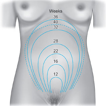
- Measure the distance abdominally from the top of the symphysis pubis over the curve of the abdomen to the top of the uterine fundus.
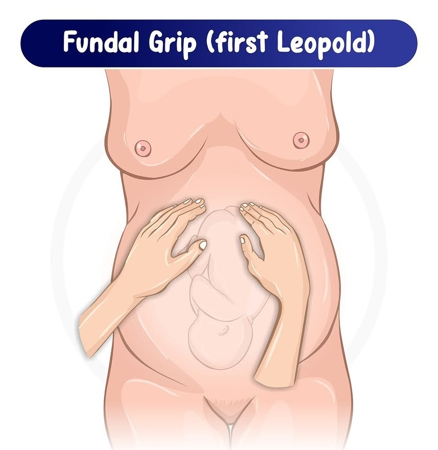
First Maneuver (Fundal Palpation)
Facing the client’s head part, palpate the fundus with both hands and observe for shape, consistency, and mobility.
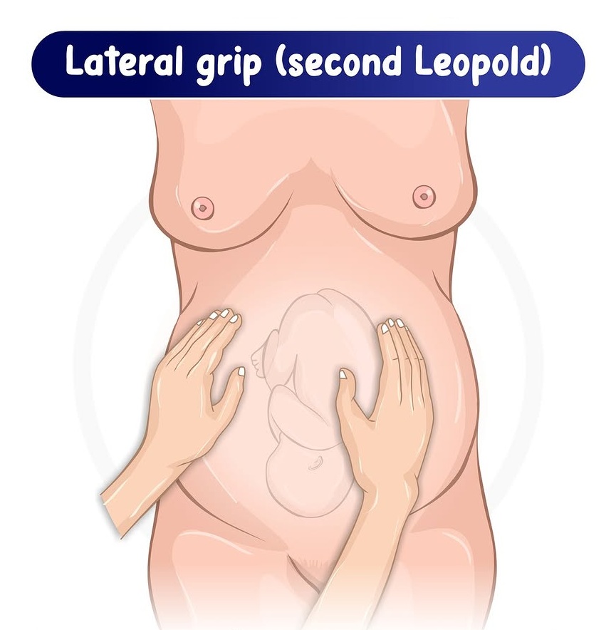
Second Maneuver (Lateral Palpation)
Still facing the client’s head part, place the left hand stationary on the right side of the uterus to immobilize it. Palpate the left side of the uterus from top to bottom with the right hand.
Apply gentle but deep pressure to determine what lies on either side of the uterus.
Repeat the procedure on the opposite side of the uterus.
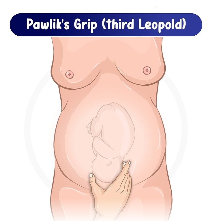
Third Maneuver (Pawlik’s Grip)
Still facing the client’s head part, grasp the lower abdomen above the symphysis pubis with the hand nearer the mother’s legs using thumbs and fingers.
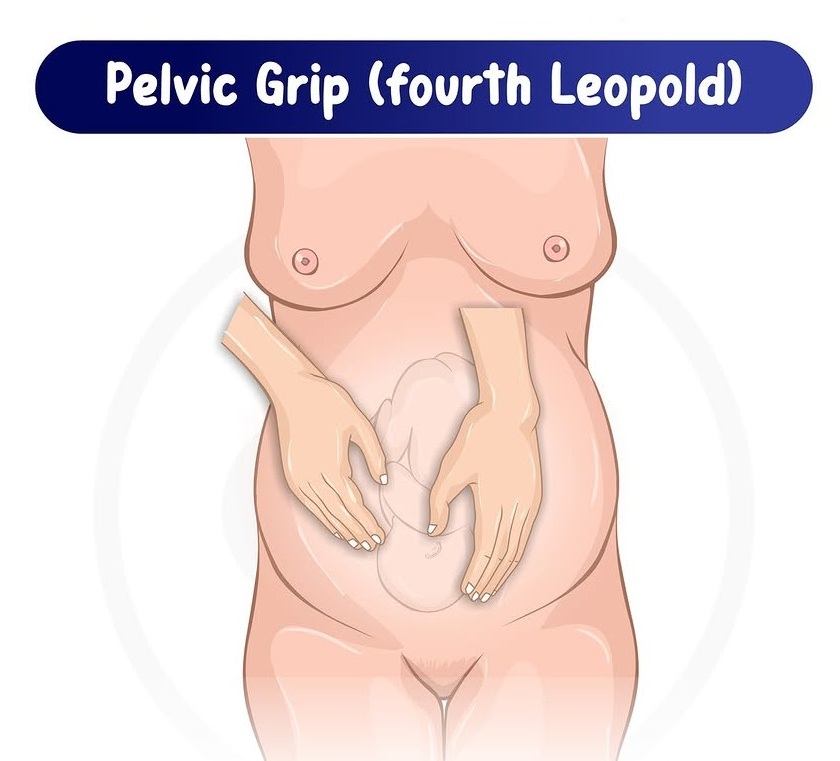
Fourth Maneuver (Pelvic Palpation)
Facing the client’s foot part, place the tips of the first three fingers on both sides of the lower uterus about 2 inches above the inguinal region.
Press downward and inward in the direction of the birth canal. Note that the fingers on one hand meet no obstruction and glide over the nape of the baby’s neck. The other hand meets an obstruction, which is the brow of the baby.
Auscultate the Fetal Heart Tone
- Remember the fetal back from the second maneuver.
- Place the bell of the stethoscope at the area of the fetal back.
- Count for 1 full minute while comparing with the mother’s pulse.
Place the patient in a comfortable position.
Relay and interpret the findings to the mother.
EVALUATION
Evaluate client comfort and patient’s response to the procedure.
DOCUMENTATION
Record the findings correctly.